Search
PATENT PORTFOLIO
Health, Medicine, and Biotechnology
NASA's portfolio of health, medical, and biotechnology is a testament to the Agency's innovation. From developing advanced materials for use in space suits to creating innovative medical devices and treatments, NASA has a long history of advancing the field of health and medicine. In this portfolio, you will find a diverse range of patents that have been developed by NASA scientists and engineers that can enable new healthcare technologies.

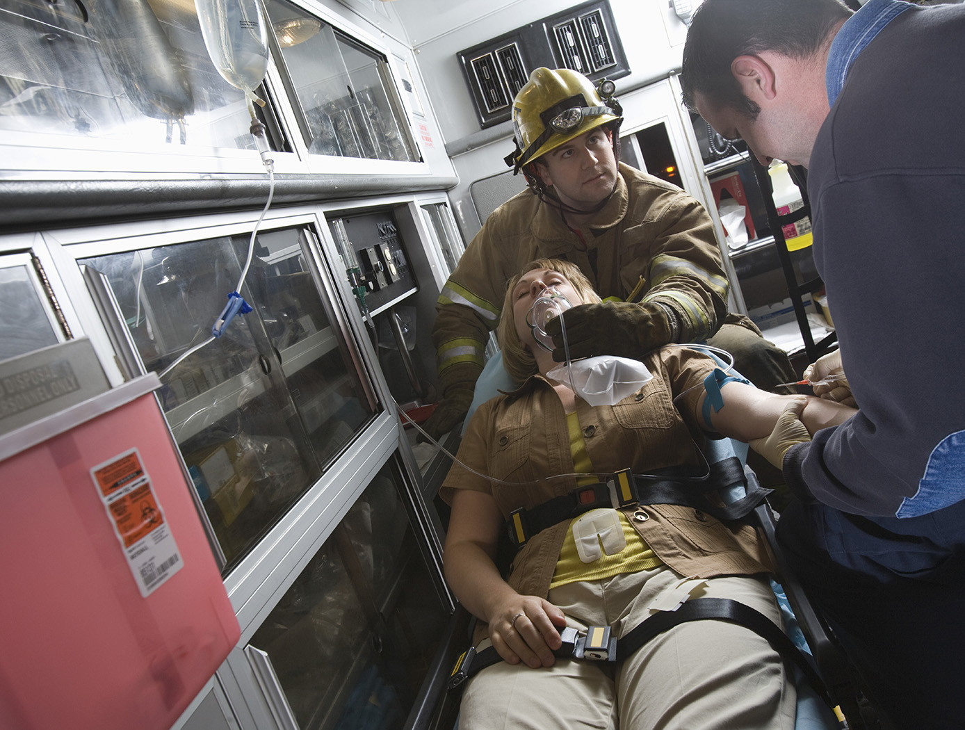
Solid And Liquid Waste Drying Bag
This invention addresses the problem of human solid waste disposal in microgravity, and consists of a soft-sided container or bag that (1) collects wet material using airflow, (2) compacts material under vacuum, and (3) dries material under applied vacuum. End products are clean water and dried, compacted, and bagged material. The bag includes a liquid-impermeable and vapor-impermeable outer layer and a liquid-impermeable but vapor-permeable inner layer membrane, defining an inner bag, through which some vapor can pass. The port is located in the outer layer, and activation of the vacuum source causes some of the original vapors and vaporized liquids to pass through the membrane liner. Liquid components of the moist waste solids within the bag may also be vaporized and transported across the membrane. Waste solids, such as excrement, remain in an inner layer defined by the membrane, and are partly dried by withdrawal of vaporized liquid and vaporized liquid components in the moist solids. These waste solids are thereby trapped and sealable in the bag, while the original vapors and the vaporized portion of the liquids pass through the membrane and are received by an outer bag defined by the membrane and the outer layer of the bag. After use, the bag is sealed and stored for ultimate disposal.

HeartBeatID
Cardiac muscle is myogenic and is capable of generating an action potential and depolarizing and repolarizing signals from within the muscle itself. An intrinsic conduction system (ICS), a group of specialized cardiac cells, passes an electrical signal throughout the heart. This technology is a method and associated system to identify a person based on the use of statistical parameters, peak amplitudes and/or time interval lengths and/or depolarization-repolarization vector angles and/or depolarization-repolarization vector lengths for PQRST electrical signals associated with heart waves. The statistical parameters, estimated to be at least 192, serve as biometric indicia to authenticate or to decline to authenticate an asserted identity of a candidate person. There are three on-line modes of operation enrollment, verification, and identification as well as two off-line modes statistics and settings. In enrollment the raw electrocardiography (ECG) signal is processed and the results in the form of parameters are serialized and saved. Verification and Identification procedures use the feature parameters for recognition (classification) of subjects based on the same kind of parameters (features) of heartbeats extracted from the ECG signal of a person to be verified or identified.

Miniature Bioreactor System for Cell Culture
The miniature bioreactor system was developed to provide the capabilities for NASA to perform cell studies in space and then provide results back to investigators on Earth with minimal tools and cost. The miniature bioreactor system has the potential to also be used on Earth as a laboratory bench-top cell culturing system without the need for expensive equipment and reagents.
The system can be operated under computer control to reduce the operator handling and to reduce result variations. The system includes a bioreactor, a fluid-handling subsystem, a chamber wherein the bioreactor is maintained in a controlled atmosphere and temperature, and control subsystems. The system can be used to culture both anchorage dependent and suspension cells (prokaryotic or eukaryotic cell types). Cells can be cultured for extended periods of time in this system, and samples of cells can be extracted and analyzed at specified intervals. The miniature bioreactor system for cell culturing has applications in pharmaceutical drug screening and cell culture studies.
Subcutaneous Structure Imager
Current subcutaneous vessel imagers use large, multiple, and often separate assemblies with complicated optics to image subcutaneous structures as two-dimensional maps on a wide monitor, or as maps extracted by a computer and focused onto the skin by a video projection. The scattering of infrared light that takes place during this process produces images that are shadowy and distorted. By contrast, Glenn's innovative approach offers a relatively compact and inexpensive alternative to the conventional setup, while also producing clearer images that can be rendered in either two or three dimensions. Glenn's device uses off-the-shelf near-infrared technology that is not affected by melanin content and can also operate in dark environments.
In Glenn's novel subcutaneous imager, a camera is configured to generate a video frame. Connected to the camera is a processor that receives the signal for the video frame and adjusts the thresholds for darkness and whiteness. The result is that the vein (or other subcutaneous structure) will show very dark, while other surrounding features (which would register as gray) become closer to white due to the heightened contrast between thresholds. With no interval of complex algorithms required, the image is presented in real-time on a display, yielding immediate results. Glenn's advanced technology also allows the operator to achieve increased depth perception through the synchronization of a pair of imaging devices. Additionally, the novel use of a virtual-reality headset affords a three-dimensional view of the field, thereby improving the visualization of veins. In short, Glenn's researchers have produced an inexpensive, lightweight, high-utility device for locating and identifying subcutaneous structures in patients.

Electroactive Material for Wound Healing
An electroactive device is applied to an external wound site. This method utilizes generated low level electrical stimulation to promote the wound healing process while simultaneously protecting it from infection. The material is fabricated from polyvinylidene fluoride, or PVDF, a thermoplastic fluoropolymer that is highly piezoelectric when poled. The fabrication method of the electroactive material is based on a previous Langley invention of an apparatus that is used to electrospin highly aligned polymer fiber material. A description of the fabrication method can be found in the technology opportunity announcement titled "NASA Langley's Highly Electrospun Fibers and Mats," which is available on NASA Langley's Technology Gateway.

Nanosensor Array for Medical Diagnoses
Many diseases are accompanied by characteristic odors. Their recognition can provide diagnostic clues, guide the laboratory evaluation, and affect the choice of immediate therapy. The study of the chemical composition of human breath using gas chromatography mass spectrometry (GC/MS) has shown a correlation between the volatile compounds and the occurrence of certain illnesses. The presence of those specific compounds can provide an indication of physiological malfunction and support the diagnosis of diseases. This condition requires an analytical tool with very high sensitivity for its measurement. A number of volatile compounds, so called biomarkers, are found in breath samples, normally at low parts per billion (ppb) levels. For example, the acetone in the exhaled breath from human with other biomarkers can indicate Type I diabetes. Usually, the concentration of the volatile compounds in human breath is very low and the background relative humidity is high, almost 100%. NASAs invention utilizes an array of chemical sensors combined with humidity, temperature, and pressure for real-time breath measurement to correlate the chemical information in the breath with the state and functioning of different human organs. This tool provides a non-invasive method for fast and accurate diagnosis at the medical point of care or at home. The sensor chip includes multisensors for a comprehensive measurement of chemical composition, temperature, humidity, and pressure/flow rate. The sensor data collected from this chip can be wired or wirelessly transmitted to a computer terminal at the doctors desk or hospital monitoring center. The sensor chip can be connected directly or via Universal serial bus (USB) to a cell phone for data transmission over a long distance and receive an instruction from a doctors office for an immediate therapy.

3D Construction of Biologically Derived Materials
Once genes for a desired material type, delivery mode, control method and affinity have been chosen, assembling the genetic components and creating the cell lines can be done with well-established synthetic biology techniques. A 3D microdeposition system is used to make a 3D array of these cells in a precise, microstructure pattern and shape.
The engineered cells are suspended in a printable 'ink'. The 3D microdeposition system deposits minute droplets of the cells onto a substrates surface in a designed print pattern. Additional printer passes thicken the material. The cell array is fed nutrients and reagents to activate the engineered genes within the cells to create and deposit the desired molecules. These molecules form the designed new material. If desired, the cells may be removed by flushing. The end product is thus a 3D composite microstructure comprising the novel material.
This innovation provides a fast, controlled production of natural, synthetic, and novel biomaterials with minimum resource overhead and reduced pre- and post-processing requirements.
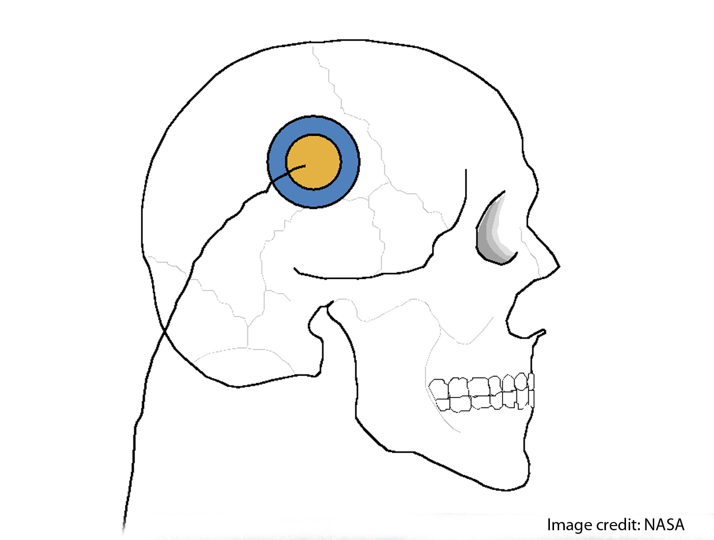
Non-invasive Intracranial Pressure Measurement
This technology and a product based on it offer new analytical capabilities for assessment of intracranial dynamics. It offers the possibility for the monitoring of transcranial expansion and related physiological phenomena in humans resulting from variations in intracranial pressure (ICP) caused by injuries to the head and/or brain pathologies. The technology uses constant frequency pulse phase-locked loop (CFPPLL) technology to measure skull expansion caused by pressure and its variations in time. This approach yields a more accurate, more robust measurement capability with improved bandwidth that allows new analytical approaches for assessing the physiology of skull expansion under pulsatile cerebral blood flow. The dynamical quantities assessable with the CFPPLL include skull volume expansion and total fluid. Such an instrument can serve to measure intracranial dynamics with equation based algorithms, and offers a path to measure or determine quasistatic intracranial pressure, along with the pulsatile related intracranial pressure increments. Supportive measurements, such as time dependence of arterial pressure waveforms together with time dependent phase change of transcranial expansions can serve as the basis of noninvasive techniques to measure intracranial pressure.

Ionic Magnetic Resonance Tailors Animal Cells/Tissues
The apparatus comprises a randomized gravity vector multiphasic culture system with a self-feeding growth module, an optionally disposable nutrient module, and a removable AIMR chamber that delivers a pulsating multivariant field to the contents of the culture system. It produces overlapping or fluctuating alternating ionic magnetic resonance frequencies at one or more modal intervals ranging from about 7.8 Hz to about 59.9 Hz to the cell chamber. The apparatus may yield better regulation that can be manipulated to allow for increased rate of cell growth, faster differentiation, increased cell fidelity, and the induction or suppression of selective physiological genes involved in directing cellular differentiation and dedifferentiation.
The use of an AIMR field may provide a significant improvement over existing bioreactors, including pulsating electromagnetic field (PEMF) and time-variance electromagnetic field (TVEMF) cellular growth induced systems, in that AIMR incorporates the modulation of cellular transcription. The AIMR system utilizes pre-sterilized disposable modules and a removable alternating ionic magnetic resonance chamber, reducing the hazard for contamination, allowing scientists to implement physiological and homeostatic parameters similar to a naturally occurring physiological system.
View more patents

