Electroactive Scaffold
Health Medicine and Biotechnology
Electroactive Scaffold (LAR-TOPS-200)
Three-dimensional scaffold that mimics native biological environment
Overview
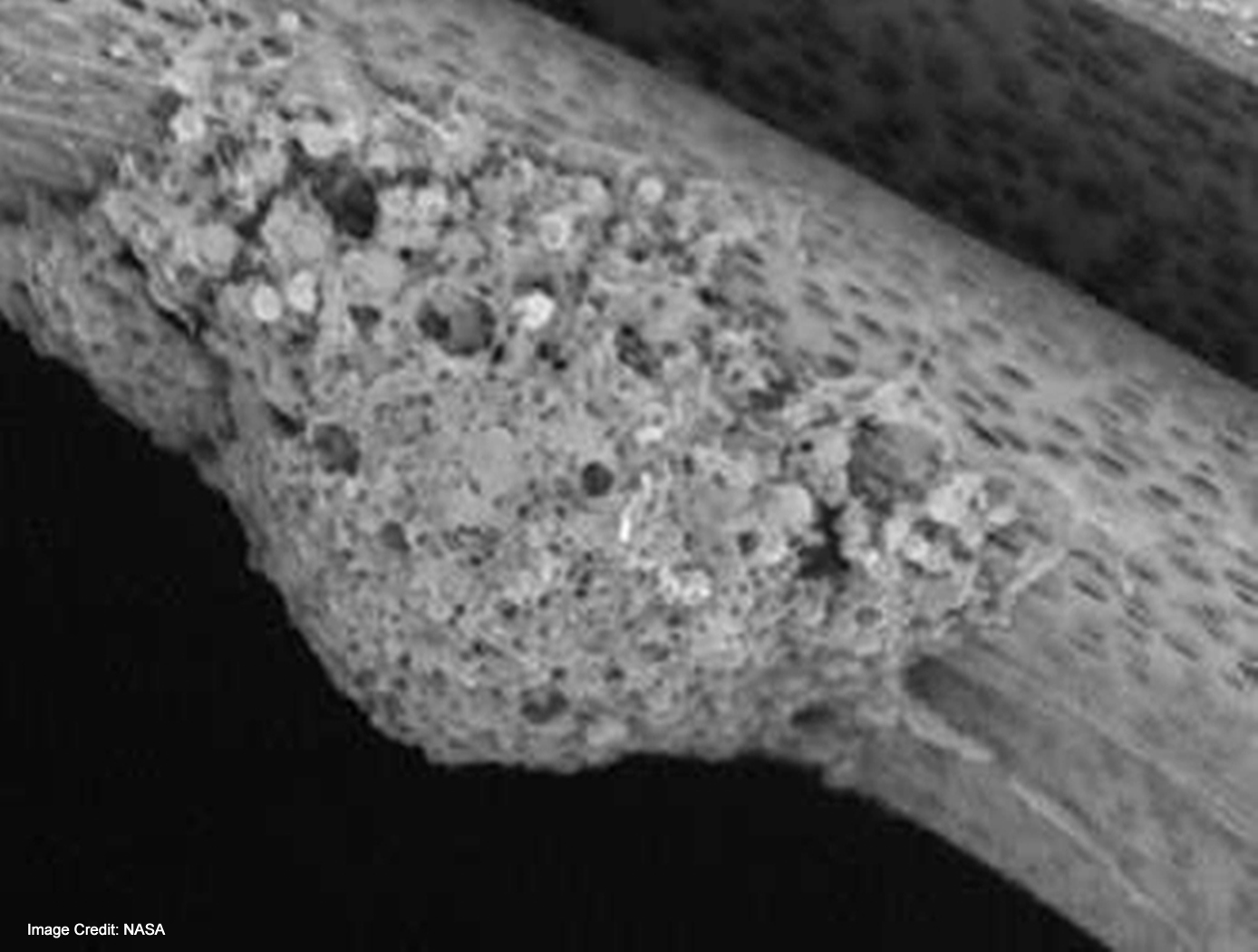
This technology, developed at NASA's Langley Research Center in collaboration with scientists at Duke University, is a novel three-dimensional scaffold structure that utilizes electroactive fibers for tissue and/or stem cell engineering. This invention enables electroactive fibers to be assembled into three-dimensional scaffolds to more closely mimic the native biological environment by providing biochemical, mechanical, and electrical cues.
The Technology
Current scaffold designs and materials do not provide all of the appropriate cues necessary to mimic in-vivo conditions for tissue engineering and stem cell engineering applications. It has been hypothesized that many biomaterials, such as bone, muscle, brain and heart tissue exhibit piezoelectric and ferroelectric properties. Typical cell seeding environments incorporate biochemical cues and more recently mechanical stimuli, however, electrical cues have just recently been incorporated in standard in-vitro examinations. In order to develop their potential further, novel scaffolds are required to provide adequate cues in the in-vitro environment to direct stem cells to differentiate down controlled pathways or develop novel tissue constructs. This invention is for a scaffold that provides for such cues by mimicking the native biological environment, including biochemical, topographical, mechanical and electrical cues.


Benefits
- Mimics the native biological environment by providing biochemical, mechanical, and electrical cues
- Can be used with adult mesenchymal stem cells
Applications
- Stem cell treatments
- Tissue engineering
- Research and development
Technology Details
Health Medicine and Biotechnology
LAR-TOPS-200
LAR-17789-1
LAR-17789-3
Similar Results

Highly Aligned Electrospun Fibers and Mats
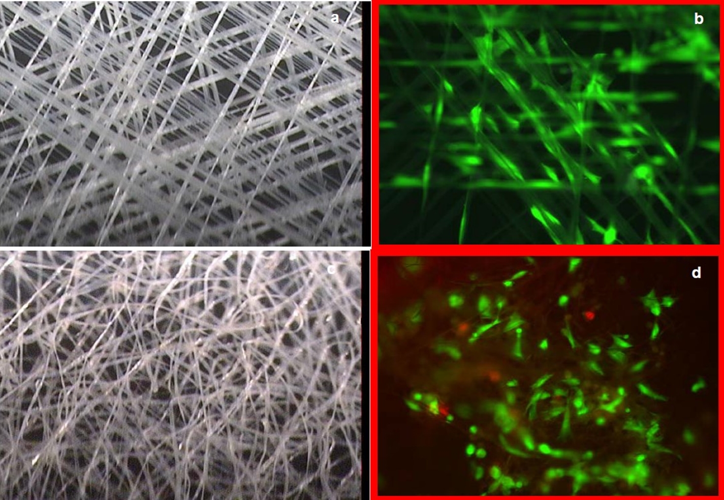
Electrospinning offers a versatile way to produce one-dimensional micro- or nanometer mats; however, electrospun fibers are typically collected in a random orientation, which limits their applications. NASA has developed a new apparatus that uses an auxiliary counter electrode to align fibers for control of the fiber distribution during the spinning process. The electrostatic force imposed by the auxiliary electrode creates a converged electric field, which affords control over the distribution of the fibers on the rotating collector surface.
The process begins when a pump slowly expels polymer solution through the tip of
the spinneret at a set flow rate as a positive charge is applied. The auxiliary electrode,
which is negatively charged, is positioned opposite the charged spinneret. The disparity
in charges creates an electric field that effectively controls the behavior of the polymer
jet as it is expelled from the spinneret; it ultimately controls the distribution of the
fibers and mats formed from the polymer solution as it lands on the rotating collection
mandrel. A broad range of fiber diameters can be manufactured by modifying various
parameters of the process and/or polymer solution. Performance data has confirmed
the substantial role that the electric field plays in the significant improvement in fiber
alignment and control relative to using the rotating collector alone.
Prototypes have been produced, and the repeatability of the process has been
confirmed. A patent application has been filed.

3D Construction of Biologically Derived Materials
Once genes for a desired material type, delivery mode, control method and affinity have been chosen, assembling the genetic components and creating the cell lines can be done with well-established synthetic biology techniques. A 3D microdeposition system is used to make a 3D array of these cells in a precise, microstructure pattern and shape.
The engineered cells are suspended in a printable 'ink'. The 3D microdeposition system deposits minute droplets of the cells onto a substrates surface in a designed print pattern. Additional printer passes thicken the material. The cell array is fed nutrients and reagents to activate the engineered genes within the cells to create and deposit the desired molecules. These molecules form the designed new material. If desired, the cells may be removed by flushing. The end product is thus a 3D composite microstructure comprising the novel material.
This innovation provides a fast, controlled production of natural, synthetic, and novel biomaterials with minimum resource overhead and reduced pre- and post-processing requirements.

Electroactive Material for Wound Healing
An electroactive device is applied to an external wound site. This method utilizes generated low level electrical stimulation to promote the wound healing process while simultaneously protecting it from infection. The material is fabricated from polyvinylidene fluoride, or PVDF, a thermoplastic fluoropolymer that is highly piezoelectric when poled. The fabrication method of the electroactive material is based on a previous Langley invention of an apparatus that is used to electrospin highly aligned polymer fiber material. A description of the fabrication method can be found in the technology opportunity announcement titled "NASA Langley's Highly Electrospun Fibers and Mats," which is available on NASA Langley's Technology Gateway.

Micro scale electro hydrodynamic (EHD) modular cartridge pump
NASA GSFCs EHD pump uses electric fields to move a dielectric fluid coolant in a thermal loop to dissipate heat generated by electrical components with a low power system. The pump has only a few key components and no moving parts, increasing the simplicity and robustness of the system. In addition, the lightweight pump consumes very little power during operation and is modular in nature. The pump design takes a modular approach to the pumping sections by means of an electrically insulating cartridge casing that houses the high voltage and ground electrodes along with spacers that act as both an insulator and flow channel for the dielectric fluid. The external electrical connections are accomplished by means of commercially available pin and jack assemblies that are configurable for a variety of application interfaces. It can be sized to work with small electric components or lab-on-a-chip devices and multiple pumps can be placed in line for pumping greater distances or used as a feeder system for smaller downstream pumps. All this is done as a one-piece construction consolidating an assembly of 21 components over previous iterations.

Ionic Magnetic Resonance Tailors Animal Cells/Tissues
The apparatus comprises a randomized gravity vector multiphasic culture system with a self-feeding growth module, an optionally disposable nutrient module, and a removable AIMR chamber that delivers a pulsating multivariant field to the contents of the culture system. It produces overlapping or fluctuating alternating ionic magnetic resonance frequencies at one or more modal intervals ranging from about 7.8 Hz to about 59.9 Hz to the cell chamber. The apparatus may yield better regulation that can be manipulated to allow for increased rate of cell growth, faster differentiation, increased cell fidelity, and the induction or suppression of selective physiological genes involved in directing cellular differentiation and dedifferentiation.
The use of an AIMR field may provide a significant improvement over existing bioreactors, including pulsating electromagnetic field (PEMF) and time-variance electromagnetic field (TVEMF) cellular growth induced systems, in that AIMR incorporates the modulation of cellular transcription. The AIMR system utilizes pre-sterilized disposable modules and a removable alternating ionic magnetic resonance chamber, reducing the hazard for contamination, allowing scientists to implement physiological and homeostatic parameters similar to a naturally occurring physiological system.


