Ultrasonic System To Assess Compartment Syndrome
health medicine and biotechnology
Ultrasonic System To Assess Compartment Syndrome (LAR-TOPS-162)
An ultrasonic means and method to assess whether Compartment Syndrome has occurred
Overview
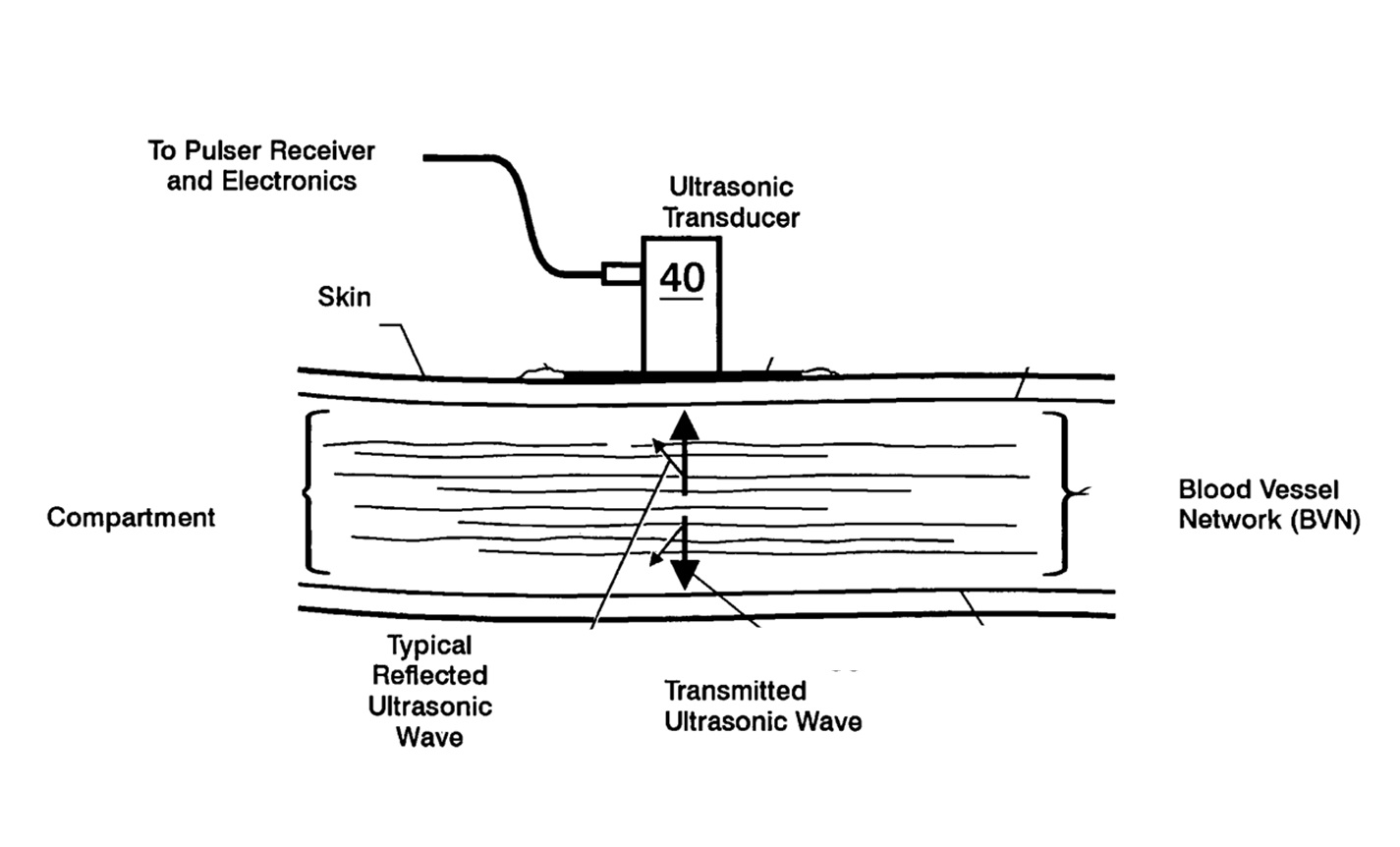
NASA Langley Research Center has developed an ultrasonic system to assess Compartment Syndrome. Compartment Syndrome occurs when bleeding or swelling interfere with proper blood circulation in enclosed groups of muscles and nerves. Most commonly this occurs after a substantial injury, such as a broken arm or leg. Without proper treatment, myoneural necrosis occurs, leading to permanent injury and possible limb amputation. Even experienced physicians can have trouble making a reliable diagnosis of Compartment Syndrome and current testing for Compartment Syndrome requires invasive procedures. This invention provides a non-invasive and quick system to test for Compartment Syndrome, and to monitor for its possible onset.
The Technology
The technology uses ultrasonic waves to categorize pressure build-up in a body compartment. The method includes assessing the body compartment configuration and identifying the effect of pulsatile components on at least one compartment dimension. An apparatus is used for measuring excess pressure in the body compartment having components for imparting ultrasonic waves such as a transducer, placing the transducer to impart the ultrasonic waves, capturing the reflected imparted ultrasonic waves, and converting them to electrical signals, a pulsed phase-locked loop device for assessing a body compartment configuration and producing an output signal, and means for mathematically manipulating the output signal to thereby categorize pressure build-up in the body compartment to the point of interference with blood flow in the compartment from the mathematical manipulations.


Benefits
- Non-invasive
- Easy-to-use
Applications
- Emergency medicine
- Combat casualty care
- Sports Injuries
Similar Results

Full Spectrum Infrasonic Stethoscope for Screening Heart, Carotid Artery, and Lung Related Diseases
Microphones and stethoscopes are regularly used by physicians to detect sounds when monitoring physiological conditions. These monitors are coupled directly to a person's body and measure in certain bandwidths either by listening or by recording the signals. The physiological processes such as respiration and cardiac activity are reflected in a different frequency bandwidth from 0.01 Hz to 500 Hz. This technology can monitor physiological conditions in the entire bandwidth range. Signals can also be wirelessly transmitted, using Bluetooth, to other recording devices at any other location.

Rapid and Verified Crimping for Critical Wiring Needs
The crimping innovations are based on traditional ultrasonic nondestructive evaluation methods. The quality of the contact between the connector and wire is determined by sending an acoustic wave through the crimp assembly. As the applied pressure increases and the crimp terminal deforms around the wire, the ultrasonic signature passing through the crimp is altered. The system analyzes the changes in the signal, including the amplitude and frequency content, as an indication of the quality of both the electrical and mechanical connection between the wire and terminal. Various crimp quality issues such as undercrimping, missing wire strands, incomplete wire insertion, partial insulation removal, and incorrect wire gauge have been tested using this technique, and results show that the instrumented crimp tool consistently discriminates between good and poor crimps for all of these potential quality issues. This information can be used to provide a pass or fail indication for instant verification of the crimp quality and to give a better prediction for the service life of the crimp.

Subcutaneous Structure Imager
Current subcutaneous vessel imagers use large, multiple, and often separate assemblies with complicated optics to image subcutaneous structures as two-dimensional maps on a wide monitor, or as maps extracted by a computer and focused onto the skin by a video projection. The scattering of infrared light that takes place during this process produces images that are shadowy and distorted. By contrast, Glenn's innovative approach offers a relatively compact and inexpensive alternative to the conventional setup, while also producing clearer images that can be rendered in either two or three dimensions. Glenn's device uses off-the-shelf near-infrared technology that is not affected by melanin content and can also operate in dark environments.
In Glenn's novel subcutaneous imager, a camera is configured to generate a video frame. Connected to the camera is a processor that receives the signal for the video frame and adjusts the thresholds for darkness and whiteness. The result is that the vein (or other subcutaneous structure) will show very dark, while other surrounding features (which would register as gray) become closer to white due to the heightened contrast between thresholds. With no interval of complex algorithms required, the image is presented in real-time on a display, yielding immediate results. Glenn's advanced technology also allows the operator to achieve increased depth perception through the synchronization of a pair of imaging devices. Additionally, the novel use of a virtual-reality headset affords a three-dimensional view of the field, thereby improving the visualization of veins. In short, Glenn's researchers have produced an inexpensive, lightweight, high-utility device for locating and identifying subcutaneous structures in patients.

System for In-situ Defect Detection in Composites During Cure
NASA's System for In-situ Defect (e.g., porosity, fiber waviness) Detection in Composites During Cure consists of an ultrasonic portable automated C-Scan system with an attached ultrasonic contact probe. This scanner is placed inside of an insulated vessel that protects the temperature-sensitive components of the scanner. A liquid nitrogen cooling systems keeps the interior of the vessel below 38°C. A motorized X-Y raster scanner is mounted inside an unsealed cooling container made of porous insulation boards with a cantilever scanning arm protruding out of the cooling container through a slot. The cooling container that houses the X-Y raster scanner is periodically cooled using a liquid nitrogen (LN2) delivery system. Flexible bellows in the slot opening of the box minimize heat transfer between the box and the external autoclave environment. The box and scanning arm are located on a precision cast tool plate. A thin layer of ultrasonic couplant is placed between the transducer and the tool plate. The composite parts are vacuum bagged on the other side of the tool plate and inspected. The scanning system inside of the vessel is connected to the controller outside of the autoclave. The system can provide A-scan, B-scan, and C-scan images of the composite panel at multiple times during the cure process.
The in-situ system provides higher resolution data to find, characterize, and track defects during cure better than other cure monitoring techniques. In addition, this system also shows the through-thickness location of any composite manufacturing defects during cure with real-time localization and tracking. This has been demonstrated for both intentionally introduced porosity (i.e., trapped during layup) as well processing induced porosity (e.g., resulting from uneven pressure distribution on a part). The technology can be used as a non-destructive evaluation system when making composite parts in in an oven or an autoclave, including thermosets, thermoplastics, composite laminates, high-temperature resins, and ceramics.

Electroactive Material for Wound Healing
An electroactive device is applied to an external wound site. This method utilizes generated low level electrical stimulation to promote the wound healing process while simultaneously protecting it from infection. The material is fabricated from polyvinylidene fluoride, or PVDF, a thermoplastic fluoropolymer that is highly piezoelectric when poled. The fabrication method of the electroactive material is based on a previous Langley invention of an apparatus that is used to electrospin highly aligned polymer fiber material. A description of the fabrication method can be found in the technology opportunity announcement titled "NASA Langley's Highly Electrospun Fibers and Mats," which is available on NASA Langley's Technology Gateway.


