Electrochemical Sensors Based on Enzyme-Linked Immunosorbent Assay
Health Medicine and Biotechnology
Electrochemical Sensors Based on Enzyme-Linked Immunosorbent Assay (TOP2-307)
Electrochemical ELISA Microelectrode Array Biosensor
Overview
Biological assays are ever evolving to move towards lower limits of detection and improved sensitivity. Improvements in trace biological molecule detection can have significant impact in healthcare, food safety and environmental safety industries. Detection of trace biological molecules can be critical to the diagnosis of early onset of diseases or infections. Researchers at NASA Ames Research Center developed an electrochemical, bead-based biological sensor based on Enzyme-Linked Immunosorbent Assay (ELISA) combining a magnetic concentration of signaling molecules and electrochemical amplification using wafer-scale fabrication of microelectrode arrays. Originally developed for the detection of the SARS-CoV-2 nucleoprotein, this invention can be easily modified to detect other environmental or human health biomarkers.
The Technology
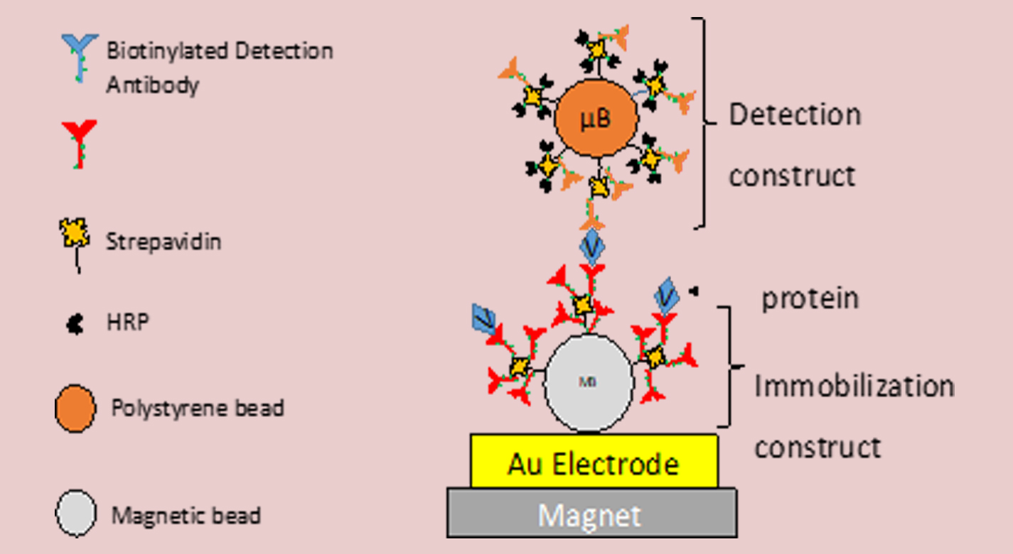
NASA’s electrochemical Enzyme-Linked Immunosorbent Assay (ELISA) microelectrode array biosensor advantageously incorporates a microbead detection construct, coupled with a magnetic immobilization construct, which substantially increases the signal sensitivity of a sensor. The magnetic immobilization construct draws the microbead detection construct to an electrode detection surface, enhancing signal sensitivity. By concentrating the signaling molecules close to the electrode detection surface, electrochemical redox cycling is achieved by reducing the distance between the two, allowing for regeneration of reporter molecules.
Whereas a traditional ELISA testing exhibits five to ten signaling molecules per probe molecule binding event, the present electrochemical ELISA-based biosensor testing exhibits up to 4,857 signaling molecules per probe molecule binding event. The model bead construct exhibits a more than 6.75-fold in increased measured signal, and more than 35.7-fold improvement in signal sensitivity. When compared to traditional optical ELISA, the present invention improves the limit of detection by up to a factor of 60.5.
NASA’s electromagnetic ELISA-based biosensor can be used for the detection of SARS-CoV-2 virus to enhance Covid-19 testing during the early phases of infection. The technology may also be modified to detect other biomarkers.

Benefits
- Trace biological molecule detection
- 6.75-fold increased measured signal
- 35.7-fold increased signal sensitivity
- Simultaneous detection of two or more biological molecules
- Magnetic concentration and enrichment of electrochemical signal
Applications
- Biomedical diagnostic devices market
- Electrochemical biosensors market
- Point-of-care diagnostics market
Technology Details
Health Medicine and Biotechnology
TOP2-307
ARC-18692-1
|
Tags:
|
Similar Results

Systems and methods employing nanomaterial sensors for detecting conditions impacting a Volatile Organic Compounds (VOCs) profile in breath
The technology involves a sophisticated system designed to detect conditions through the analysis of exhaled breath, utilizing an array of nanomaterial sensors fabricated upon a standard printed circuit board with interdigitated electrodes. These sensors are configured to interact with a sample gas that contains various Volatile Organic Compounds (VOCs) associated with a variety of biological conditions. Each sensor consists of nanomaterials, such as carbon nanotubes, composite nanotubes, nanoparticle-doped nanotubes, or polymer-coated nanotubes, all disposed on an electrically conductive structure. These sensors are highly sensitive to specific VOCs at a broad spectrum of concentrations, and each sensor generates a unique measurable electrical signal on interaction with VOCs in the breath that reflects the presence and concentration of specific components in the sample gas. The previously nanosensor diagnosis technology has been further developed to identify 64 specific formulations of nanomaterials that exhibit unique and varying sensitivities to VOCs, which enables unique response signatures to be developed for a wide range of VOCs. A single device may be developed using these principles to detect a variety of health conditions and diseases.

Biomarker Sensor Arrays for Microfluidics Applications
This invention provides a method and system for fabricating a biomarker sensor array by dispensing one or more entities using a precisely positioned, electrically biased nanoprobe immersed in a buffered fluid over a transparent substrate. Fine patterning of the substrate can be achieved by positioning and selectively biasing the probe in a particular region, changing the pH in a sharp, localized volume of fluid less than 100 nm in diameter, resulting in a selective processing of that region. One example of the implementation of this technique is related to Dip-Pen Nanolithography (DPN), where an Atomic Force Microscope probe can be used as a pen to write protein and DNA Aptamer inks on a transparent substrate functionalized with silane-based self-assembled monolayers. But it would be recognized that the invention has a much broader range of applicability. For example, the invention can be applied to formation of patterns using biological materials, chemical materials, metals, polymers, semiconductors, small molecules, organic and inorganic thins films, or any combination of these.

Inexpensive Microsensor Fabrication Process
Because chemical sensors are used in many aspects of space missions, NASA researchers are continually developing ever smaller and more robust sensors that can be manufactured inexpensively and in high quantities; e.g., in batches. Glenn has developed a way to inexpensively fabricate microsensors using a sacrificial template approach. A nanostructure, such as a carbon nanotube, serves as a template, which can then be coated with a high-temperature oxide material. The carbon nanotube can be burned off, or sacrificed, leaving only the metal oxide. The resulting structure provides the unique morphology and properties of the carbon nanotube, which are advantageous for sensing, along with the material durability and high-temperature sensing capabilities of the metal oxide. This technique increases the surface area available for sensing because both the interior and exterior of the resulting microsensor can be used for gas detection, significantly increasing performance.
The fabrication of these microsensors includes three major steps: (1) synthesis of the porous metal or metal oxide nanostructures using a sacrificial template, (2) deposition of the electrodes onto alumina substrates, and (3) alignment of the nanostructures between the electrodes. The invention has been demonstrated for methane detection at room temperature (using tin oxide, with carbon nanotubes as the sacrificial template). The microsensor offers low power consumption (no heating required), compact size, extremely low cost, and simple batch-fabrication.

Micro-Organ Device Mimics Organ Structures for Lab Testing
The MOD platform technology represents a small, lightweight, and reproducible in vitro drug screening model that could inexpensively mimic different mammalian tissues for a multitude of applications. The technology is automated and imposes minimal demands for resources (power, analytes, and fluids). The MOD technology uses titanium isopropoxide to bond a microscale support to a substrate and uses biopatterning and 3D tissue bioprinting on a microfluidic microchip to eliminate variations in local seeding density while minimizing selection pressure. With the MOD, pharmaceutical companies can test more candidates and concentrate on those with more promise therefore, reducing R&D overall cost.
This innovation overcomes major disadvantages of conventional in vitro and in vivo experimentation for purposes of investigating effects of medicines, toxins, and possibly other foreign substances. For example, the MOD platform technology could host life-like miniature assemblies of human cells and the effects observed in tests performed could potentially be extrapolated more readily to humans than could effects observed in conventional in vivo cell cultures, making it possible to reduce or eliminate experimentation on animals.
The automated NASA developed technology with minimal footprint and power requirements, micro-volumes of fluids and waste, high throughput and parallel analyses on the same chip, could advance the research and development for new drugs and materials.

Completely biodegradable filtration system for waste metal recovery from aqueous solution
There is a significant need for an inexpensive biological approach to recover specific, targeted metals and other target materials in e-waste or other aqueous solutions that requires minimal input of resources, including energy. This invention is a method of removing or adsorbing a target substance or material, for example, a metal, non-metal toxin, dye, or small molecule drug, from solution, by functionalizing a substrate with a peptide configured to selectively bind to the target substance or material and to bind to the substrate. The substrate is fungal mycelium, and the naturally-occurring or bioengineered peptide is called a target-binding domain, which is chemically bonded to a selected solid substrate. The target chemical species binds to the target-binding domain and is removed from solution. The target can be any chemical species dissolved or suspended in the solution. Capture of the target by the substrate can isolate and allow removal of the target substance from solution, or for utilization in water filtration, or recovery of targeted chemical species from solution, particularly aqueous solution applications. The peptides used include (i) fusion peptides and/or proteins containing metal-binding domain sequence and optionally containing substrate-binding domain sequence; (ii) fusion peptides/proteins containing a metal-binding domain and a chitin-binding domain; and (iii) nucleic acids encoding fusion peptides and/or proteins containing metal-binding domain sequence. The technology enables simple scale up to a level that could be successfully implemented in an environment with limited resources, such as on a space mission or on earth in developing countries with poor access to clean water.


