Search
PATENT PORTFOLIO
Health, Medicine, and Biotechnology
NASA's portfolio of health, medical, and biotechnology is a testament to the Agency's innovation. From developing advanced materials for use in space suits to creating innovative medical devices and treatments, NASA has a long history of advancing the field of health and medicine. In this portfolio, you will find a diverse range of patents that have been developed by NASA scientists and engineers that can enable new healthcare technologies.

Noninvasive Therapy for Cartilage Regeneration
Research has shown that exposure of mammalian cartilage and bone tissue to tuned magnetic fields modifies genetic regulation at a cellular level. PEMF therapy relies on modulation and resonance of weak metals (ions) such as Ca2+, K+, Li+, and Mg2+ which can be made to move at the sub-cellular level when exposed to magnetic flux. This NASA technology is a device and method for modifying genetic regulation of cartilage and bone in response to PEMF therapy and may serve as the basis for development of novel therapies for cartilage diseases.
In initial studies, cultured human chondrocyte cells (HCH) from patients with early-stage osteoarthritis were exposed to PEMF stimulation using a variety of tuned electro-magnetic pulse characteristics such as flux magnitude, slew rates, rise and fall times, frequency, wavelength, and duty cycle. Waveforms used in testing were monophasic, bi-phasic, square, sinusoidal, and triangular in nature. Frequencies were generally low, ranging from 6-500 Hz, and the waveforms used high rising and falling slew rates on the order of Tesla/sec, promoting pulses or bursts.
Cellular catabolic and anabolic gene expression analyses comprised of fold-change (in expression) were accomplished by a survey of 47,000 human genes using an AFFYMETRIX Gene Array. Results show that variation of waveform used in PEMF therapies, independent of flux intensity, influences genetic regulation of HCH from patients with early-stage osteoarthritis.

Non-invasive Intracranial Pressure Measurement
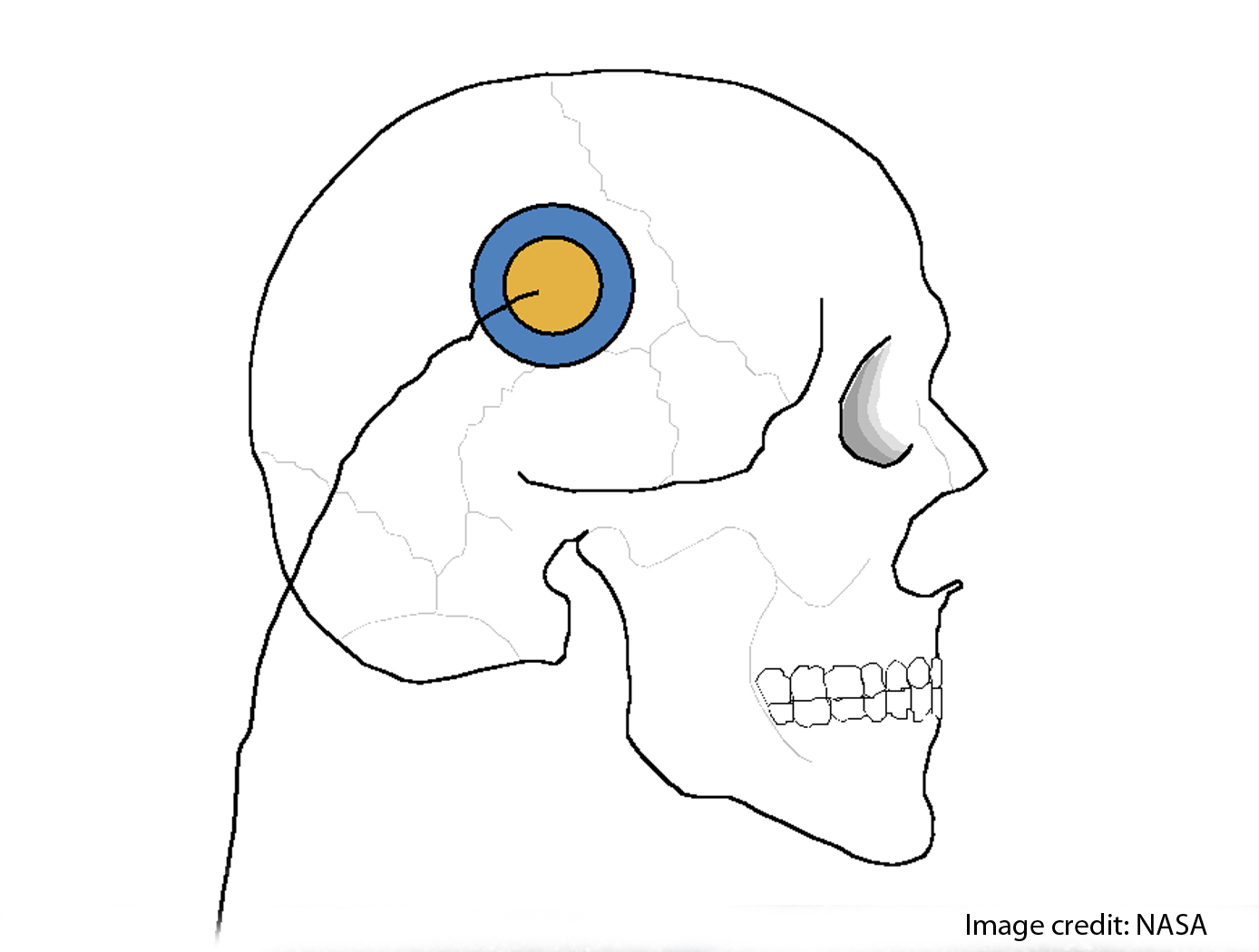
This technology and a product based on it offer new analytical capabilities for assessment of intracranial dynamics. It offers the possibility for the monitoring of transcranial expansion and related physiological phenomena in humans resulting from variations in intracranial pressure (ICP) caused by injuries to the head and/or brain pathologies. The technology uses constant frequency pulse phase-locked loop (CFPPLL) technology to measure skull expansion caused by pressure and its variations in time. This approach yields a more accurate, more robust measurement capability with improved bandwidth that allows new analytical approaches for assessing the physiology of skull expansion under pulsatile cerebral blood flow. The dynamical quantities assessable with the CFPPLL include skull volume expansion and total fluid. Such an instrument can serve to measure intracranial dynamics with equation based algorithms, and offers a path to measure or determine quasistatic intracranial pressure, along with the pulsatile related intracranial pressure increments. Supportive measurements, such as time dependence of arterial pressure waveforms together with time dependent phase change of transcranial expansions can serve as the basis of noninvasive techniques to measure intracranial pressure.

Full Spectrum Infrasonic Stethoscope for Screening Heart, Carotid Artery, and Lung Related Diseases
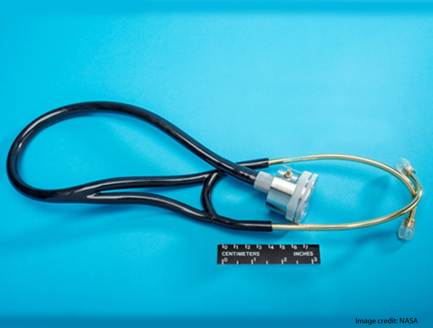
Microphones and stethoscopes are regularly used by physicians to detect sounds when monitoring physiological conditions. These monitors are coupled directly to a person's body and measure in certain bandwidths either by listening or by recording the signals. The physiological processes such as respiration and cardiac activity are reflected in a different frequency bandwidth from 0.01 Hz to 500 Hz. This technology can monitor physiological conditions in the entire bandwidth range. Signals can also be wirelessly transmitted, using Bluetooth, to other recording devices at any other location.

HeartBeatID
Cardiac muscle is myogenic and is capable of generating an action potential and depolarizing and repolarizing signals from within the muscle itself. An intrinsic conduction system (ICS), a group of specialized cardiac cells, passes an electrical signal throughout the heart. This technology is a method and associated system to identify a person based on the use of statistical parameters, peak amplitudes and/or time interval lengths and/or depolarization-repolarization vector angles and/or depolarization-repolarization vector lengths for PQRST electrical signals associated with heart waves. The statistical parameters, estimated to be at least 192, serve as biometric indicia to authenticate or to decline to authenticate an asserted identity of a candidate person. There are three on-line modes of operation enrollment, verification, and identification as well as two off-line modes statistics and settings. In enrollment the raw electrocardiography (ECG) signal is processed and the results in the form of parameters are serialized and saved. Verification and Identification procedures use the feature parameters for recognition (classification) of subjects based on the same kind of parameters (features) of heartbeats extracted from the ECG signal of a person to be verified or identified.

Oculometric Testing for Detecting/Characterizing Mild Neural Impairment
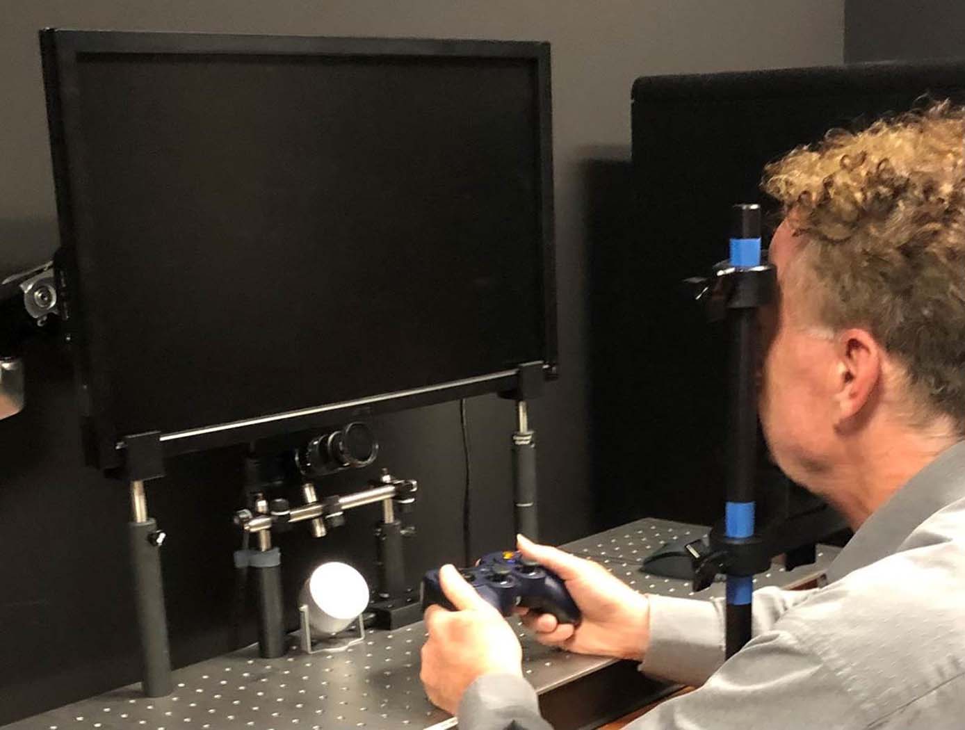
To assess various aspects of dynamic visual and visuomotor function including peripheral attention, spatial localization, perceptual motion processing, and oculomotor responsiveness, NASA developed a simple five-minute clinically relevant test that measures and computes more than a dozen largely independent eye-movement-based (oculometric) measures of human neural performance. This set of oculomotor metrics provide valid and reliable measures of dynamic visual performance and may prove to be a useful assessment tool for mild functional neural impairments across a wide range of etiologies and brain regions. The technology may be useful to clinicians to localize affected brain regions following trauma, degenerative disease, or aging, to characterize and quantify clinical deficits, to monitor recovery of function after injury, and to detect operationally-relevant altered or impaired visual performance at subclinical levels. This novel system can be used as a sensitive screening tool by comparing the oculometric measures of an individual to a normal baseline population, or from the same individual before and after exposure to a potentially harmful event (e.g., a boxing match, football game, combat tour, extended work schedule with sleep disruption, blast or toxic exposure, space mission), or on an ongoing basis to monitor performance for recovery to baseline. The technology provides set of largely independent metrics of visual and visuomotor function that are sensitive and reliable within and across observers, yielding a signature multidimensional impairment vector that can be used to characterize the nature of a mild deficit, not just simply detect it. Initial results from peer-reviewed studies of Traumatic Brain Injury, sleep deprivation with and without caffeine, and low-dose alcohol consumption have shown that this NASA technology can be used to assess subtle deficits in brain function before overt clinical symptoms become obvious, as well as the efficacy of countermeasures.

Apparatus and Method for Biofeedback Training
Measured values of physiological signals may be associated with physiological states and may be used to define the presence of such states. For example, in a physiological state of anxiety, adrenaline diverts blood from the body surface to the core of the body in response to a perceived danger. As warm blood is withdrawn from the surface of the skin, the skin temperature drops. Similarly, in a physiological state of stress, perspiration generally increases making the skin more conductive to the passage of an electrical current, thereby increasing the galvanic skin response.
It is well known in the field of performance psychology that the peak performance of a task, such as, for example, putting in golf, foul shooting in basketball, serving in tennis, marksmanship in archery or on a gunnery range, shooting pool, or throwing darts, requires the presence of a physiological state, comprising one or more optimal measured values of physiological signals, coincident with the physical performance of the task. The presence of such an optimal physiological state in athletics is colloquially referred to as being in the zone.
The technology provides:
an apparatus and method of performance-enhancing biofeedback training that has intuitive and motivational appeal to the trainee, by tightly embedding the biofeedback training in the actual task whose performance is to be improved; and,
an apparatus and method of performance-enhancing biofeedback training that is operational in real-time, precisely at the moment when a task or exercise, such as an athletic or military maneuver, is required to be performed.
The feedback behavior of the physical environment provided by the present invention has the added benefit of providing aids to visualization that the trainee can use in the real-world skill performance setting.

Electroactive Material for Wound Healing
An electroactive device is applied to an external wound site. This method utilizes generated low level electrical stimulation to promote the wound healing process while simultaneously protecting it from infection. The material is fabricated from polyvinylidene fluoride, or PVDF, a thermoplastic fluoropolymer that is highly piezoelectric when poled. The fabrication method of the electroactive material is based on a previous Langley invention of an apparatus that is used to electrospin highly aligned polymer fiber material. A description of the fabrication method can be found in the technology opportunity announcement titled "NASA Langley's Highly Electrospun Fibers and Mats," which is available on NASA Langley's Technology Gateway.

Subcutaneous Structure Imager
Current subcutaneous vessel imagers use large, multiple, and often separate assemblies with complicated optics to image subcutaneous structures as two-dimensional maps on a wide monitor, or as maps extracted by a computer and focused onto the skin by a video projection. The scattering of infrared light that takes place during this process produces images that are shadowy and distorted. By contrast, Glenn's innovative approach offers a relatively compact and inexpensive alternative to the conventional setup, while also producing clearer images that can be rendered in either two or three dimensions. Glenn's device uses off-the-shelf near-infrared technology that is not affected by melanin content and can also operate in dark environments.
In Glenn's novel subcutaneous imager, a camera is configured to generate a video frame. Connected to the camera is a processor that receives the signal for the video frame and adjusts the thresholds for darkness and whiteness. The result is that the vein (or other subcutaneous structure) will show very dark, while other surrounding features (which would register as gray) become closer to white due to the heightened contrast between thresholds. With no interval of complex algorithms required, the image is presented in real-time on a display, yielding immediate results. Glenn's advanced technology also allows the operator to achieve increased depth perception through the synchronization of a pair of imaging devices. Additionally, the novel use of a virtual-reality headset affords a three-dimensional view of the field, thereby improving the visualization of veins. In short, Glenn's researchers have produced an inexpensive, lightweight, high-utility device for locating and identifying subcutaneous structures in patients.

System for Incorporating Physiological Self-Regulation Challenge into Parcourse/Orienteering Type Games and Simulations
Although biofeedback is an effective treatment for various physiological problems and can be used to optimize physiological functioning in many ways, the benefits can only be attained through a number of training sessions, and such gains can only be maintained over time through regular practice. However, adherence to regular training has been a problem that has plagued the field of physiological self-regulation limiting its utility. As with any exercise, incorporating biofeedback training with another activity encourages participation and enhances its usefulness.
View more patents


